51 results
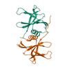
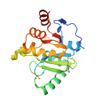
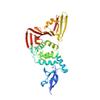
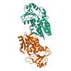
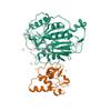
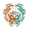
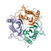
X-ray diffraction data for the Structure of IDP01693/yjeA, a potential t-RNA synthetase from Salmonella typhimurium
First author:
W.W. Navarre
Gene name: yjeA
Resolution: 1.95 Å
R/Rfree: 0.18/0.24
Gene name: yjeA
Resolution: 1.95 Å
R/Rfree: 0.18/0.24
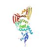
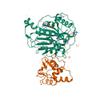
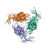
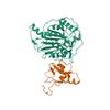
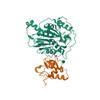
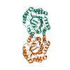
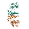
X-ray diffraction data for the Crystal Structure of NSP15 Endoribonuclease from SARS CoV-2.
First author:
Y. Kim
Gene name: orf1ab
Resolution: 2.20 Å
R/Rfree: 0.16/0.18
Gene name: orf1ab
Resolution: 2.20 Å
R/Rfree: 0.16/0.18
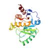
X-ray diffraction data for the The crystal structure of Papain-Like Protease of SARS CoV-2 , P3221 space group
First author:
J. Osipiuk
Gene name: orf1ab
Resolution: 1.79 Å
R/Rfree: 0.16/0.17
Gene name: orf1ab
Resolution: 1.79 Å
R/Rfree: 0.16/0.17
X-ray diffraction data for the Structure of IDP00107, a potential N-acetyl-gamma-glutamylphosphate reductase from Shigella flexneri

First author:
A.U. Singer
Gene name: argC
Resolution: 2.00 Å
R/Rfree: 0.17/0.22
Gene name: argC
Resolution: 2.00 Å
R/Rfree: 0.17/0.22
X-ray diffraction data for the The crystal structure of Nsp9 RNA binding protein of SARS CoV-2
First author:
K. Tan
Gene name: orf1ab
Resolution: 2.95 Å
R/Rfree: 0.24/0.28
Gene name: orf1ab
Resolution: 2.95 Å
R/Rfree: 0.24/0.28
X-ray diffraction data for the The crystal structure of Papain-Like Protease of SARS CoV-2 , C111S mutant
First author:
J. Osipiuk
Gene name: orf1ab
Resolution: 1.60 Å
R/Rfree: 0.12/0.16
Gene name: orf1ab
Resolution: 1.60 Å
R/Rfree: 0.12/0.16
X-ray diffraction data for the Crystal structure of the 3-oxoacyl-acyl carrier protein reductase, FabG, from Staphylococcus aureus
First author:
S.M. Anderson
Gene name: fabG
Resolution: 1.90 Å
R/Rfree: 0.16/0.19
Gene name: fabG
Resolution: 1.90 Å
R/Rfree: 0.16/0.19
X-ray diffraction data for the Crystal Structure of ADP ribose phosphatase of NSP3 from SARS CoV-2 in the complex with ADP ribose
First author:
K. Michalska
Gene name: orf1ab
Resolution: 1.50 Å
R/Rfree: 0.15/0.17
Gene name: orf1ab
Resolution: 1.50 Å
R/Rfree: 0.15/0.17
X-ray diffraction data for the Crystal Structure of ADP ribose phosphatase of NSP3 from SARS-CoV-2 in complex with MES
First author:
K. Michalska
Gene name: orf1ab
Resolution: 1.07 Å
R/Rfree: 0.13/0.15
Gene name: orf1ab
Resolution: 1.07 Å
R/Rfree: 0.13/0.15
X-ray diffraction data for the Crystal structure of type I 3-dehydroquinate dehydratase (aroD) from Clostridium difficile with covalent reaction intermediate
First author:
S.H. Light
Gene name: aroD
Resolution: 2.20 Å
R/Rfree: 0.19/0.24
Gene name: aroD
Resolution: 2.20 Å
R/Rfree: 0.19/0.24
X-ray diffraction data for the Crystal Structure of ADP ribose phosphatase of NSP3 from SARS CoV-2
First author:
Y. Kim
Gene name: orf1ab
Resolution: 2.03 Å
R/Rfree: 0.19/0.23
Gene name: orf1ab
Resolution: 2.03 Å
R/Rfree: 0.19/0.23
X-ray diffraction data for the Crystal Structure of Nsp16-Nsp10 from SARS-CoV-2 in Complex with 7-methyl-GpppA and S-Adenosylmethionine.
First author:
G. Minasov
Gene name: orf1ab
Resolution: 2.00 Å
R/Rfree: 0.16/0.18
Gene name: orf1ab
Resolution: 2.00 Å
R/Rfree: 0.16/0.18
X-ray diffraction data for the The crystal structure of papain-like protease of SARS CoV-2
First author:
J. Osipiuk
Gene name: orf1ab
Resolution: 2.70 Å
R/Rfree: 0.24/0.31
Gene name: orf1ab
Resolution: 2.70 Å
R/Rfree: 0.24/0.31
X-ray diffraction data for the Crystal Structure of ADP ribose phosphatase of NSP3 from SARS-CoV-2 in the apo form
First author:
K. Michalska
Gene name: orf1ab
Resolution: 1.35 Å
R/Rfree: 0.10/0.14
Gene name: orf1ab
Resolution: 1.35 Å
R/Rfree: 0.10/0.14
X-ray diffraction data for the The crystal structure of Papain-Like Protease of SARS CoV-2 , C111S mutant, at room temperature
First author:
J. Osipiuk
Gene name: orf1ab
Resolution: 2.48 Å
R/Rfree: 0.15/0.19
Gene name: orf1ab
Resolution: 2.48 Å
R/Rfree: 0.15/0.19
X-ray diffraction data for the 1.98 Angstrom Resolution Crystal Structure of NSP16-NSP10 Heterodimer from SARS-CoV-2 in Complex with Sinefungin
First author:
G. Minasov
Gene name: orf1ab
Resolution: 1.98 Å
R/Rfree: 0.16/0.18
Gene name: orf1ab
Resolution: 1.98 Å
R/Rfree: 0.16/0.18
X-ray diffraction data for the 2.0 Angstrom Resolution Crystal Structure of Nsp16-Nsp10 Heterodimer from SARS-CoV-2 in Complex with S-Adenosyl-L-Homocysteine

First author:
G. Minasov
Gene name: orf1ab
Resolution: 2.00 Å
R/Rfree: 0.17/0.19
Gene name: orf1ab
Resolution: 2.00 Å
R/Rfree: 0.17/0.19
X-ray diffraction data for the 1.5 Angstrom Crystal Structure of Shikimate Dehydrogenase 1 from Peptoclostridium difficile.
First author:
G. Minasov
Gene name: aroE
Resolution: 1.50 Å
R/Rfree: 0.16/0.19
Gene name: aroE
Resolution: 1.50 Å
R/Rfree: 0.16/0.19
X-ray diffraction data for the 1.80 Angstrom Resolution Crystal Structure of NSP16 - NSP10 Complex from SARS-CoV-2
First author:
G. Minasov
Gene name: orf1ab
Resolution: 1.80 Å
R/Rfree: 0.15/0.16
Gene name: orf1ab
Resolution: 1.80 Å
R/Rfree: 0.15/0.16
X-ray diffraction data for the 1.95 Angstrom Resolution Crystal Structure of NSP10 - NSP16 Complex from SARS-CoV-2
First author:
G. Minasov
Gene name: orf1ab
Resolution: 1.95 Å
R/Rfree: 0.16/0.18
Gene name: orf1ab
Resolution: 1.95 Å
R/Rfree: 0.16/0.18
X-ray diffraction data for the The 1.9 A Crystal Structure of NSP15 Endoribonuclease from SARS CoV-2 in the Complex with a Citrate
First author:
Y. Kim
Gene name: orf1ab
Resolution: 1.90 Å
R/Rfree: 0.16/0.19
Gene name: orf1ab
Resolution: 1.90 Å
R/Rfree: 0.16/0.19
X-ray diffraction data for the Crystal Structure of Nsp16-Nsp10 Heterodimer from SARS-CoV-2 in Complex with 7-methyl-GpppA and S-adenosyl-L-homocysteine.
First author:
G. Minasov
Gene name: orf1ab
Resolution: 2.10 Å
R/Rfree: 0.17/0.19
Gene name: orf1ab
Resolution: 2.10 Å
R/Rfree: 0.17/0.19
X-ray diffraction data for the 2.35 Angstrom resolution crystal structure of a putative tRNA (guanine-7-)-methyltransferase (trmD) from Staphylococcus aureus subsp. aureus MRSA252
First author:
A.S. Halavaty
Gene name: trmD
Resolution: 2.35 Å
R/Rfree: 0.21/0.26
Gene name: trmD
Resolution: 2.35 Å
R/Rfree: 0.21/0.26
X-ray diffraction data for the Crystal Structure of Nsp16-Nsp10 Heterodimer from SARS-CoV-2 with 7-methyl-GpppA and S-adenosyl-L-homocysteine in the Active Site and Sulfates in the mRNA Binding Groove.
First author:
G. Minasov
Gene name: orf1ab
Resolution: 2.25 Å
R/Rfree: 0.16/0.19
Gene name: orf1ab
Resolution: 2.25 Å
R/Rfree: 0.16/0.19
X-ray diffraction data for the 1.95 Angstrom Crystal Structure of of Type I 3-Dehydroquinate Dehydratase (aroD) from Clostridium difficile with Covalent Modified Comenic Acid.
First author:
G. Minasov
Gene name: aroD
Resolution: 1.95 Å
R/Rfree: 0.16/0.20
Gene name: aroD
Resolution: 1.95 Å
R/Rfree: 0.16/0.20
X-ray diffraction data for the Crystal structure of YqeH GTPase from Bacillus anthracis with dGDP bound
First author:
J.S. Brunzelle
Gene name: YqeH
Resolution: 1.80 Å
R/Rfree: 0.22/0.24
Gene name: YqeH
Resolution: 1.80 Å
R/Rfree: 0.22/0.24
X-ray diffraction data for the Type II citrate synthase from Vibrio vulnificus.
First author:
J. Osipiuk
Gene name: gltA
Resolution: 2.50 Å
R/Rfree: 0.19/0.23
Gene name: gltA
Resolution: 2.50 Å
R/Rfree: 0.19/0.23
X-ray diffraction data for the Crystal structure of unliganded anabolic ornithine carbamoyltransferase from Vibrio vulnificus at 1.86 A resolution
First author:
I.G. Shabalin
Resolution: 1.86 Å
R/Rfree: 0.16/0.20
Resolution: 1.86 Å
R/Rfree: 0.16/0.20
X-ray diffraction data for the 2.8 Angstrom Crystal Structure of Type III Secretion Protein YscO from Vibrio parahaemolyticus
First author:
G. Minasov
Resolution: 2.80 Å
R/Rfree: 0.19/0.24
Resolution: 2.80 Å
R/Rfree: 0.19/0.24
X-ray diffraction data for the Crystal structure of anabolic ornithine carbamoyltransferase from Vibrio vulnificus in complex with carbamoyl phosphate
First author:
I.G. Shabalin
Resolution: 2.17 Å
R/Rfree: 0.17/0.21
Resolution: 2.17 Å
R/Rfree: 0.17/0.21
X-ray diffraction data for the The crystal structure of 50S ribosomal protein L25 from Vibrio vulnificus CMCP6
First author:
K. Tan
Resolution: 2.35 Å
R/Rfree: 0.21/0.25
Resolution: 2.35 Å
R/Rfree: 0.21/0.25
X-ray diffraction data for the Crystal Structure of the methyltransferase-stimulatory factor complex of NSP16 and NSP10 from SARS CoV-2.
First author:
Y. Kim
Gene name: orf1ab
Resolution: 2.00 Å
R/Rfree: 0.17/0.19
Gene name: orf1ab
Resolution: 2.00 Å
R/Rfree: 0.17/0.19
X-ray diffraction data for the Potassium transporter peripheral membrane component (trkA) from Vibrio vulnificus

First author:
E.V. Filippova
Gene name: trkA
Resolution: 2.09 Å
R/Rfree: 0.18/0.23
Gene name: trkA
Resolution: 2.09 Å
R/Rfree: 0.18/0.23
X-ray diffraction data for the Crystal structure of anabolic ornithine carbamoyltransferase from Vibrio vulnificus in complex with citrulline and inorganic phosphate
First author:
I.G. Shabalin
Resolution: 2.08 Å
R/Rfree: 0.16/0.19
Resolution: 2.08 Å
R/Rfree: 0.16/0.19
X-ray diffraction data for the Crystal structure of anabolic ornithine carbamoyltransferase from Vibrio vulnificus in complex with carbamoylphosphate and arginine
First author:
I.G. Shabalin
Resolution: 1.85 Å
R/Rfree: 0.15/0.19
Resolution: 1.85 Å
R/Rfree: 0.15/0.19
X-ray diffraction data for the Crystal structure of anabolic ornithine carbamoyltransferase from Vibrio vulnificus in complex with carbamoyl phosphate and L-norvaline
First author:
I.G. Shabalin
Resolution: 1.70 Å
R/Rfree: 0.14/0.17
Resolution: 1.70 Å
R/Rfree: 0.14/0.17
X-ray diffraction data for the 1.75 Angstrom Crystal Structure of Transcriptional Regulator ftom Vibrio vulnificus.
First author:
G. Minasov
Resolution: 1.75 Å
R/Rfree: 0.18/0.22
Resolution: 1.75 Å
R/Rfree: 0.18/0.22
X-ray diffraction data for the Crystal Structure of RRSP, a MARTX Toxin Effector Domain from Vibrio vulnificus CMCP6

First author:
G. Minasov
Resolution: 3.45 Å
R/Rfree: 0.23/0.25
Resolution: 3.45 Å
R/Rfree: 0.23/0.25
X-ray diffraction data for the 1.60 Angstrom resolution crystal structure of an arginine repressor from Vibrio vulnificus CMCP6
First author:
A.S. Halavaty
Resolution: 1.60 Å
R/Rfree: 0.18/0.20
Resolution: 1.60 Å
R/Rfree: 0.18/0.20
X-ray diffraction data for the 2.9 Angstrom Crystal Structure of Ornithine Carbamoyltransferase (ArgF) from Vibrio vulnificus
First author:
G. Minasov
Resolution: 2.91 Å
R/Rfree: 0.16/0.20
Resolution: 2.91 Å
R/Rfree: 0.16/0.20
X-ray diffraction data for the 2.17 Angstrom resolution crystal structure of malate dehydrogenase from Vibrio vulnificus CMCP6
First author:
A.S. Halavaty
Resolution: 2.17 Å
R/Rfree: 0.16/0.21
Resolution: 2.17 Å
R/Rfree: 0.16/0.21
X-ray diffraction data for the 1.95 Angstrom Resolution Crystal Structure of N-acetyl-D-glucosamine kinase from Vibrio vulnificus.
First author:
G. Minasov
Gene name: None
Resolution: 1.95 Å
R/Rfree: 0.17/0.20
Gene name: None
Resolution: 1.95 Å
R/Rfree: 0.17/0.20
X-ray diffraction data for the 2.7 Angstrom Crystal Structure of ABC transporter ATPase from Vibrio vulnificus in Complex with Adenylyl-imidodiphosphate (AMP-PNP)

First author:
G. Minasov
Resolution: 2.70 Å
R/Rfree: 0.19/0.26
Resolution: 2.70 Å
R/Rfree: 0.19/0.26
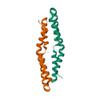
X-ray diffraction data for the 1.55 Angstrom Crystal Structure of the Four Helical Bundle Membrane Localization Domain (4HBM) of the Vibrio vulnificus MARTX Effector Domain DUF5
First author:
G. Minasov
Resolution: 1.55 Å
R/Rfree: 0.17/0.22
Resolution: 1.55 Å
R/Rfree: 0.17/0.22
X-ray diffraction data for the 1.80 Angstrom resolution crystal structure of a putative translation initiation inhibitor from Vibrio vulnificus CMCP6
First author:
A.S. Halavaty
Resolution: 1.80 Å
R/Rfree: 0.16/0.18
Resolution: 1.80 Å
R/Rfree: 0.16/0.18
X-ray diffraction data for the 2.35 Angstrom Crystal Structure of Conserved Hypothetical Protein from Toxoplasma gondii ME49.
First author:
G. Minasov
Resolution: 2.35 Å
R/Rfree: 0.20/0.26
Resolution: 2.35 Å
R/Rfree: 0.20/0.26
X-ray diffraction data for the Crystal Structure of the Catalytic Domain of the Inosine Monophosphate Dehydrogenase from Mycobacterium tuberculosis in the complex with XMP and NAD
First author:
M. Makowska-Grzyska
Gene name: guaB2
Resolution: 1.60 Å
R/Rfree: 0.16/0.19
Gene name: guaB2
Resolution: 1.60 Å
R/Rfree: 0.16/0.19
X-ray diffraction data for the Crystal Structure of the Catalytic Domain of the Inosine Monophosphate Dehydrogenase from Mycobacterium tuberculosis in the complex with IMP and the inhibitor Q67
First author:
M. Makowska-Grzyska
Gene name: guaB2
Resolution: 1.76 Å
R/Rfree: 0.15/0.18
Gene name: guaB2
Resolution: 1.76 Å
R/Rfree: 0.15/0.18
X-ray diffraction data for the Crystal Structure of the Catalytic Domain of the Inosine Monophosphate Dehydrogenase from Mycobacterium tuberculosis in the complex with IMP and the inhibitor MAD1
First author:
M. Makowska-Grzyska
Gene name: guaB2
Resolution: 1.90 Å
R/Rfree: 0.14/0.19
Gene name: guaB2
Resolution: 1.90 Å
R/Rfree: 0.14/0.19
X-ray diffraction data for the Crystal Structure of the Catalytic Domain of the Inosine Monophosphate Dehydrogenase from Mycobacterium tuberculosis in the complex with IMP and the inhibitor P41
First author:
M. Makowska-Grzyska
Gene name: guaB2
Resolution: 2.00 Å
R/Rfree: 0.18/0.22
Gene name: guaB2
Resolution: 2.00 Å
R/Rfree: 0.18/0.22
X-ray diffraction data for the Crystal Structure of the Catalytic Domain of the Inosine Monophosphate Dehydrogenase from Mycobacterium tuberculosis

First author:
M. Makowska-Grzyska
Gene name: guaB2
Resolution: 1.69 Å
R/Rfree: 0.15/0.18
Gene name: guaB2
Resolution: 1.69 Å
R/Rfree: 0.15/0.18